A thoracoscope is an endoscope for viewing the chest cavity. Physicians use it either to examine patients or to perform video-assisted thoracic surgery (VATS) – the former is medical thoracoscopy, the latter surgical thoracoscopy. The earliest known thoracoscopy took place in Dublin in 1865, and in 1910 the technique was improved by a Swedish physician.
Features of medical thoracoscopy
Medical thoracoscopy is simpler and quicker than surgical thoracoscopy. It can be performed by a physician in the bronchoscopy suite without an OR. Procedures are relatively simple and mainly diagnostic.
Features of surgical thoracoscopy: in the last century, limited technology meant chest surgery could only be performed by thoracotomy, with incisions usually 20 to 30 cm or larger and a jack-like retractor to spread the chest wall. This was extremely traumatic, recovery took a long time, and patients suffered many complications and after-effects.
In the 1980s, video display systems, miniature cameras, cold light sources and fiber optic bundles were introduced to thoracoscopy, making it easier and more accurate. In the 1990s, the USA and France each performed their first video-assisted thoracoscopic operations: using only two or three small incisions of about 1 cm in the chest wall, a video scope and miniature instruments were introduced into the chest, and the operation was performed with special instruments.
This approach avoids opening the chest: the wounds are small and the ribs need not be spread, preserving the integrity of the chest wall and the patient's breathing function and greatly increasing surgical success. Today thoracoscopy is widely used for diagnosis and treatment of chest diseases as well as lung, esophageal and mediastinal diseases – a very wide range of uses.
Thoracoscopy is considered one of the great breakthroughs in thoracic surgery of the last century and is the hallmark of minimally invasive chest surgery. Compared with conventional surgery, VATS makes higher and stricter demands on surgeons.
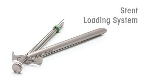
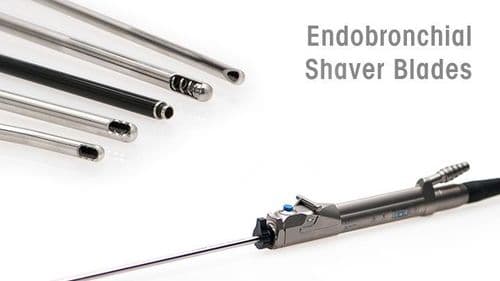
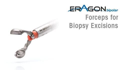
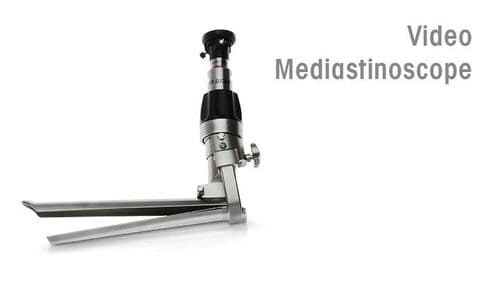
Medical instruments and equipment from Richard Wolf
Richard Wolf's unique mini thoracoscopy set for single-port technique helps physicians diagnose and treat the chest cavity. Minimally invasive diagnosis needs only a very small incision, and the extremely atraumatic approach also reduces patients' pain.
Richard Wolf offers many VATS instrument set options, whose special ergonomic handle design ensures precision, sensitivity and safety.
In diagnosis and surgery, the endoscope becomes the physician's eyes, so a clear view is extremely important. Richard Wolf has decades of experience in flexible endoscopy, and its award-winning flexible sensor bronchoscopes not only deliver excellent image quality but also fit into very confined working areas. Diagnostic, therapeutic and ultra-thin versions are available as needed, combining design and function.
Visit Pneumology / Thoracic Surgery on the Richard Wolf website to learn more about our instruments and equipment. You are also welcome to contact us directly. For our locations in China, please visit:www.richard-wolf.cn/en/contact/sales-and-service-network/.